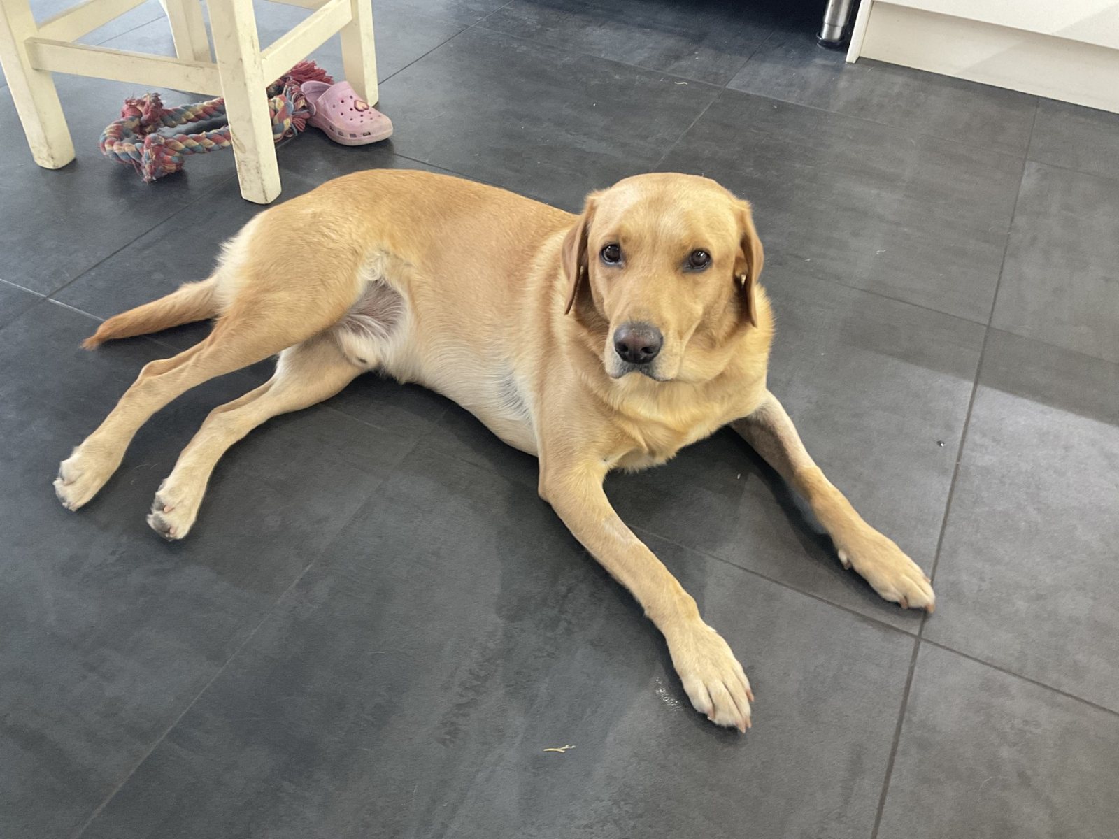
Five-year-old Labrador Teddie loves being active, but when a niggling limp wouldn't go away, a CT scan revealed a hidden weakness in his elbow.
For a few months, Teddie’s owners had noticed that he would intermittently limp on one of his front legs. It was particularly noticeable first thing in the morning and after swimming, an activity most Labradors would be very reluctant to give up!
Although the lameness would come and go, its persistence suggested there was an underlying problem. On examination, we suspected Teddie’s discomfort was coming from his elbow, so we decided it was time to take a closer look.
We performed a CT scan of Teddie’s elbows. CT uses X-rays taken from lots of different angles to build up highly detailed images of the bones and joints, allowing us to identify abnormalities that can sometimes be difficult to see on standard X-rays.
Teddie’s scan revealed a humeral intracondylar fissure, also referred to as HIF. The humerus is the large bone of the upper front leg, and its lower end forms part of the elbow joint. In Teddie’s case, there was a fine line of weakness running through this part of the bone.
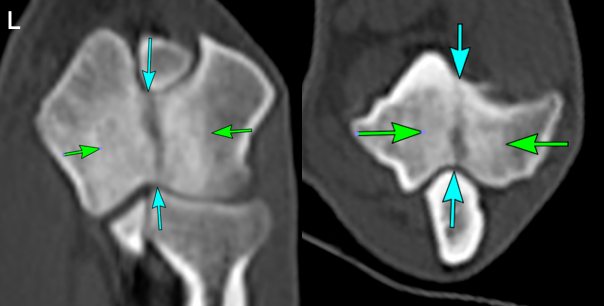
A HIF can cause ongoing elbow pain and intermittent lameness, but there is another important concern. The fissure creates a weak point in the humerus, increasing the risk that the bone could completely fracture, sometimes following relatively minor trauma.
After discussing the options with Teddie’s owners, we decided that surgery offered the best way of making his elbow more comfortable while also protecting the bone for the future.
Positioning the screw for this surgery requires considerable accuracy. It needs to pass through a relatively small area of bone in exactly the right direction while avoiding the elbow joint and other important structures.
This is where we were able to put Teddie’s CT scan to another use. Using his CT images, a patient-specific 3D-printed surgical guide was created to fit precisely onto his bone. This allowed us to plan the ideal path for the screw before we even entered the operating theatre and then reproduce that position accurately during surgery.
Vets Kyra and Cliff performed Teddie’s operation here at Bicester Vets. Using the custom guide, we drilled across the lower part of his humerus before placing a strong metal screw across the fissure. The screw stabilises the two sides of the bone, helping to reduce discomfort while also protecting the weakened humerus from suffering a complete fracture.
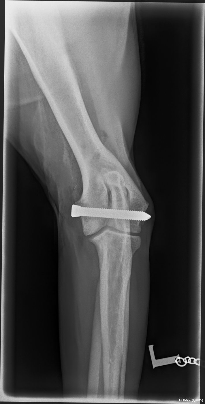
We are pleased to say Teddie is doing really well following his surgery. His lameness is resolving nicely and, after a period of carefully controlled exercise while his elbow recovers, we’re hopeful he’ll soon be back to full speed on his walks!
Teddie’s case is a great example of how veterinary medicine continues to advance. From identifying a tiny abnormality on his CT scan, to using those same images to plan his surgery and create a bespoke 3D-printed guide, we were able to tailor his treatment specifically to him.
Hopefully, it won’t be too long before Teddie is back to enjoying everything a five-year-old Labrador should, including plenty more swimming!